Mastocytoma u psa i kota objawy i leczenie [lek wet Krystyna Skiersinis]
Co to są mastocyty? Mastocyty to komórki wchodzące w skład układu immunologicznego. Mastocyty | Źródło: Wikipedia Obecne są w wielu tkankach, w bliskim sąsiedztwie naczyń krwionośnych. Znajdujemy je zwłaszcza w miejscach, które oddzielają środowisko zewnętrzne od wnętrza organizmu, takich jak: skóra, błona śluzowa dróg oddechowych,

Mastocytom
Cutaneous mastocytosis describes a group of disorders characterized by the presence of excessive numbers of mast cells in the skin. Patients with cutaneous mastocytosis do not fulfill diagnostic criteria for systemic mastocytosis and show no evidence of organ involvement other than the skin. Forms of cutaneous mastocytosis include three.
Mastocytoma u psa i kota objawy i leczenie [lek wet Krystyna Skiersinis]
Co to jest choroba mastocytów psów? Guzy komórek tucznych skóry u psów są guzy komórki tuczne, które są komórkami o funkcji odpornościowej. Interweniują m.in. w procesach alergicznych oraz w gojeniu się ran, przez co zawierają histaminę i heparynę.

Nevypočitatelný nepřítel psů Animed
A mastocytoma is a type of mastocytosis that presents as a solitary collection of mast cells in the skin. Mast cells are made in the bone marrow and are part of the body's immune system. These cells, once activated, release a variety of proteins and chemicals that create allergic reactions and attract other cell types to fight off infections.

Lekarz weterynarii Krystyna Skiersinis cowsierscipiszczy https
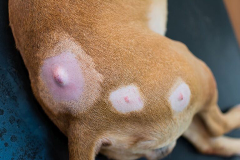
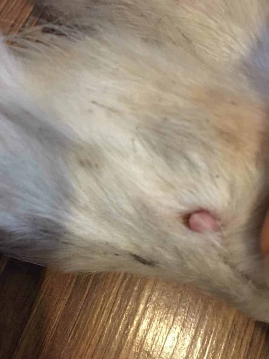
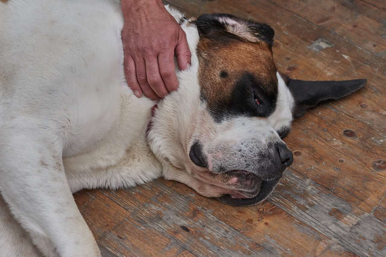
Mastocytoma u psa - rozpoznanie. Guzy komórek tucznych są znane z tego, że przypominają reakcje skórne lub wzrosty, takie jak ukąszenia owadów, brodawki i inne rodzaje guzów skóry. Dlatego ważne jest, aby skontaktować się z lekarzem weterynarii, jeśli zauważysz jakiekolwiek nieprawidłowości skórne.
Mastocitoma canino Saúde do cão e cuidados zooplus Magazine
Mastocytomas - Solitary or multiple mastocytomas of the skin are usually present at birth or develop in the first months of life . Mastocytoma lesions are similar in quality.. Mastocytosis (cutaneous and systemic) in adults: Epidemiology, pathogenesis, clinical manifestations, and diagnosis..pathologic changes in the skin that differ.
Rak skóry u psa jak rozpoznać i leczyć nowotwory skóry psów?
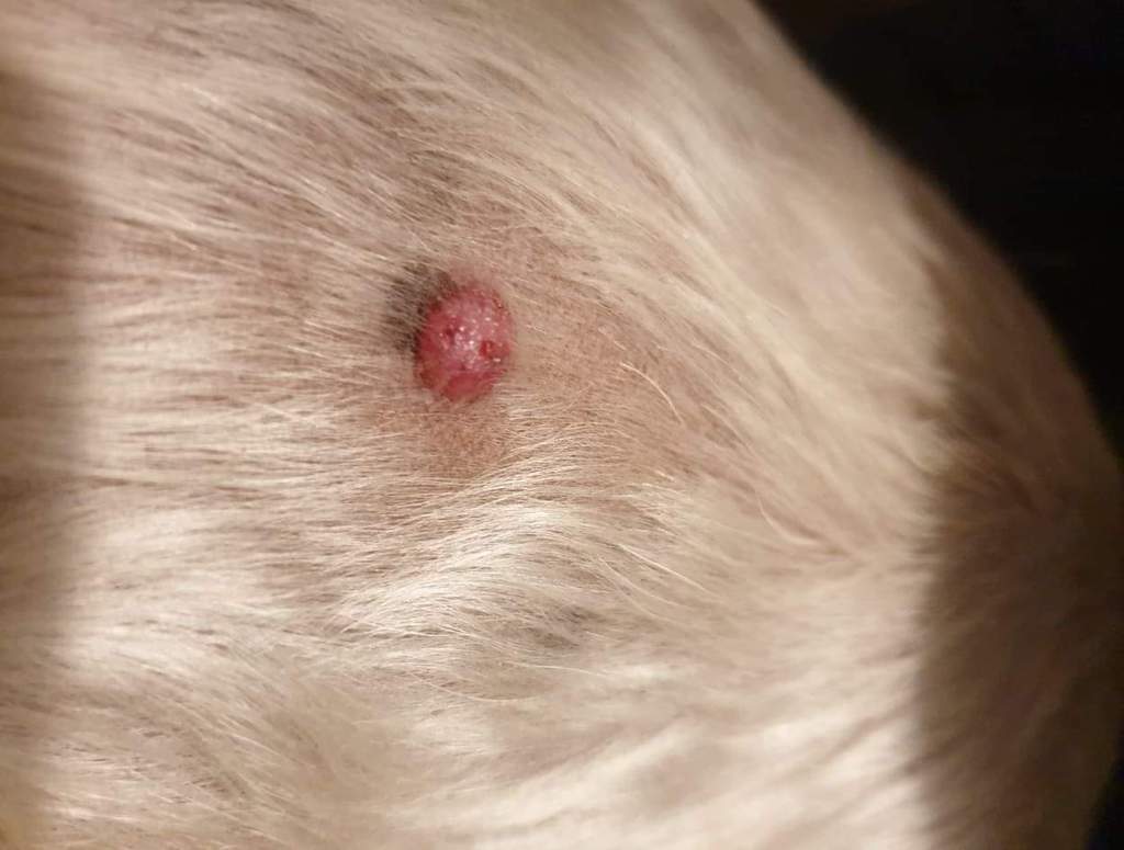
Mastocytoma u kota to natomiast najczęściej okrągłe, twarde grudki lub guzki o średnicy od kilku milimetrów do 2 cm, przeważnie bezwłose, dobrze odgraniczone od okolicznych tkanek. Tak naprawdę tylko wizyta u weterynarza i biopsja połączona z badaniem cytologicznym pozwolą rozpoznać, czy guz skóry jest niegroźny, czy też może.

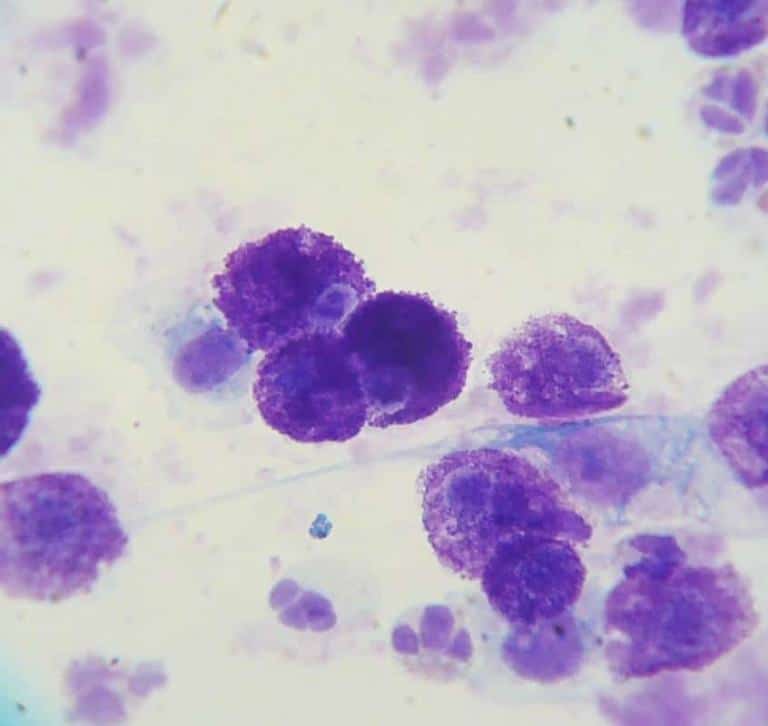
WETERYNARYJNY BLOG CYTOLOGICZNY Mastocytoma skóry w ujęciu cytologicznym.
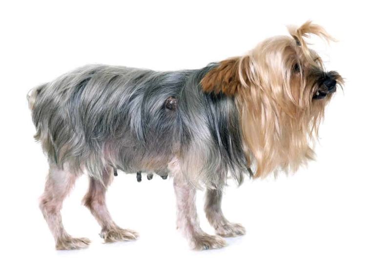
Czym jest mastocytoma u psa? Mastocytoma to nowotworowy rozrost komórek tucznych (mastocytów) znajdujących się w skórze i tkance podskórnej. Zdecydowanie rzadziej występuje postać trzewna, w której procesem nowotworowym objęte są takie narządy, jak śledziona i wątroba. Mastocytoma może wystąpić u każdego psa, jednak ryzyko jest większe w przypadku:
Mastocytoma u psa i kota groźny guz skóry Animal Center
Mast Cell Tumor (Mastocytoma) in Dogs. By Monica Tarantino, DVM on Dec. 21, 2021. In This Article. Summary. View 6 More + What are Mast Cell Tumors? Mast cell tumors are the most common form of skin cancer found in dogs. Located in connective tissue throughout the body, mast cells are white blood cells that are part of the immune system.
Mastocytoma u psa i kota objawy i leczenie [lek wet Krystyna Skiersinis]
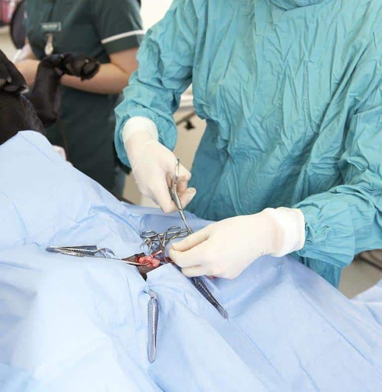
Diagnoza mastocytomy u psa opiera się na badaniu histopatologicznym, które pozwala na ocenę stopnia zaawansowania choroby. Im wcześniej rozpocznie się leczenie, tym lepsza jest prognoza dla psa. W przypadku zaawansowanych stadiów choroby, prognoza może być mniej optymistyczna, ale nie oznacza to, że nie ma szans na poprawę stanu zdrowia psa.

Nevypočitatelný nepřítel psů Animed
Prostate cancer occurs when cancer cells develop in or around the prostate. It is the most diagnosed cancer in men in the United States each year, and according to the National Cancer Institute (NCI), approximately 11.2 percent of all men in the U.S. will be diagnosed with the disease at some point during their lifetime.

Jak wygląda świerzb u psa (ZDJĘCIA) i jak leczyć? Rozpieszczony.pl
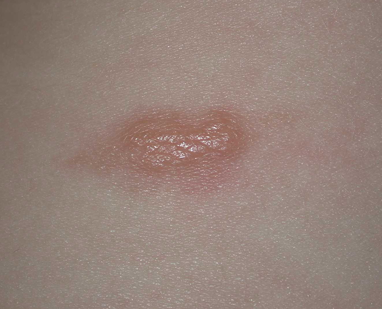
Background: The diagnosis of solitary cutaneous mastocytoma is mainly clinical, based on lesion morphology, the presence of a positive Darier sign, and the absence of systemic involve-ment. Knowledge of this condition is important so that an accurate diagnosis can be made. Objective: To familiarize physicians with the clinical manifestations, diagnosis, evaluation, and man-agement of a.
Mastocytoma u psa i kota objawy i leczenie [lek wet Krystyna Skiersinis]
Objectives: Review the etiology of mastocytoma. Describe the presentation of mastocytoma. Summarize the treatment of mastocytoma. Outline the importance of improving care coordination among interprofessional team members to improve outcomes for patients affected by mastocytoma. Access free multiple choice questions on this topic. Go to:

Mastocytoma u psa i kota objawy i leczenie [lek wet Krystyna Skiersinis]
Mastocytoma jest jednym z najczęściej występujących guzów skóry u psów. Nazywana jest kameleonem wśród nowotworów skóry. Diagnoza: Jak diagnozuje się guzy z komórek tucznych u psów? Jeśli zauważysz u psa zwiększone masy tkankowe lub inne zmiany skórne, koniecznie skonsultuj się z lekarzem weterynarii, który je dokładnie obejrzy i zbada palpacyjnie.
solitary mastocytoma pictures, photos
Guzy z komórek tucznych (mastocytoma) są jednymi z najczęstszych nowotworów skóry psów. Sprawdź jak wygląda diagnostyka i leczenie mastocytomy!
Narośl Na Skórze Psa Zdjęcia Polska Zdjecia
Introduction. Cutaneous mastocytosis can present as either urticarial pigmentosa (also referred to as maculopapular cutaneous mastocytosis), diffuse cutaneous mastocytosis, telangiectasia macularis perstans, and mastocytoma [1-5].Mastocytomas are most commonly seen in infants (under 2 years of age) and children (ranging in age from 2 year to less than 16 years); many of these lesions improve.